|
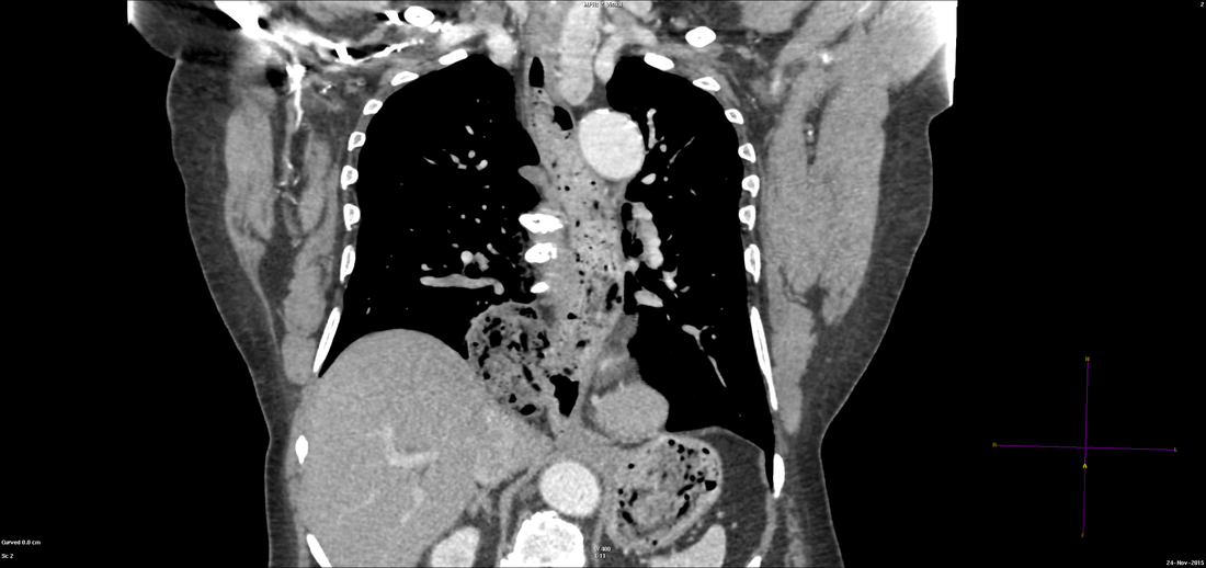
Difficulty Level: Radiology - Mid ++ (FRCR, EDiR, ABR) The axial images provided demonstrate that there is dilatation of the oesophagus with extensive food residue. Inferiorly new the diaphragm there is a focal dilatation of the oesophagus. This appearance can raise several considerations. In the first instance one may consider whether there has been a gastric pull up following lower oesophagectomy (Ivor Lewis procedure), however, there are no surgical sutures present and the stomach is below the diaphragm in its entirety. One may also consider whether there is a dysmotility of the oesophagus, such as achalasia or scleroderma. The latter is not associated with food residue but rather a patulous oesophagus, prone to reflux. The former is a consideration, however, there is insufficient dilatation just above the level of the gastro-oesophageal junction and does not account for the focal dilatation of the oesophagus. One can also consider whether the focal dilatation of the oesophagus relates to a sliding or paraesophageal hernia, however, in either of these hernias this should be two entrances/connections to the hernia from the oesophagus. One connection reflecting the inferior oesophageal continuation to the stomach, the more superior connection demonstrating the connection from the hernia to the more proximal oesophagus. In this instance, however, axial images demonstrate only a single focal connection to this dilatation of the oesophagus. Therefore, this finding reflects a diverticulum. In this typical location the findings are due to an epiphrenic diverticulum. The findings become very much more apparent on the curved planar reconstuction below. Large diverticula of the intrathoracic oesophagus can be due to traction effects, occurring historically due tuberculous nodes resulting in traction of the oesophagus in the mid oesophagus. Epiphrenic diverticula are, however, pulsion diverticula, usually arising along the right posterolateral margin of the distal oesophagus just above the gastro-oesophageal junction. These diverticula are associated with raised intraluminal pressure and therefore may be associated with distal oesophageal webs/strictures or generalised motility disorders such as achalasia. They are false diverticula in that they are focal herniations of the submucosa and mucosa through the muscularis propria. These may predispose to severe reflux, food regurgitation and aspiration. They are frequently, as in this case originally, misinterpreted as hiatus hernias. Resection is advised and may be performed by VATS/Laparoscopy.
0 Comments
|
From Grayscale
Latest news about Grayscale Courses, Cases to Ponder and other info Categories
All
Archives
October 2018
|

|
|
Grayscale Courses est. 2015

 RSS Feed
