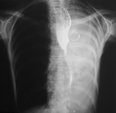
The additional film provided demonstrates the collapsed left lung, this image having been performed following visualization of the abnormalities on the images provided. In order to combine the findings into one malignancy diagnosis, the best unifying diagnosis is lung carcinoma with mediastinal extension or malignant nodal disease secondarily infiltrating the oesophagus. The reverse process is less likely, as mediastinal adenopathy from oesophageal carcinoma would be unlikely to peripherally extend into the left hilum without obvious abnormality on the right side. Lymphoma could bridge the anatomical distance between the oesophagus and the left main bronchus but such infiltrative disease of the oesophagus would be unusual.
The findings in this case were not individually difficult, however, our eyes are conditioned to observing certain findings according to the clinical indication or examination. When presented with the findings individually it is easy to reach the correct diagnosis, yet in combination they may prove more difficult. It is always important to look at the entire film on every case. For CT this would also imply visualization of all window levels and settings.


 RSS Feed
