So consider:
High atomic number pneumoconiosis (e.g.stannosis (tin) or baritosis (barium) or silicosis- look for upper lobe predominance and dense septal lines
Post lymphangiogram (not done any more)
Haemosiderosis (primary: children, secondary: look for mitral valve disease)
Alveolar microlithiasis (nodules usually a little bigger, often perivascular and may be outlined by black band of pericardial or pleural edges) and
Heterotopic pulmonary calcification (hyperparathyroidism)- for which this appearance is characteristic.
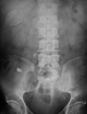
On CT these nodules are subpleural and anterior. Bone isotope imaging may show diffuse pulmonary and gastric uptake even in the absence of plain film findings. Porcelain gallbladder in same patient and calcified mesenteric nodes – postulated related to hyperparathyroidism in this patient. More characteristically an abdominal film may demonstrate nephrocalcinosis.


 RSS Feed
