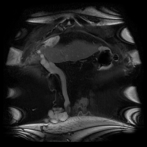
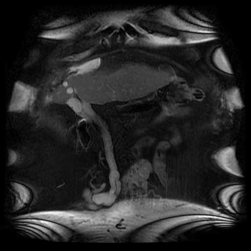
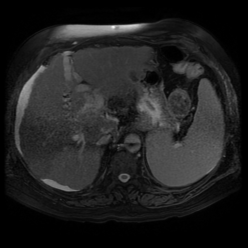
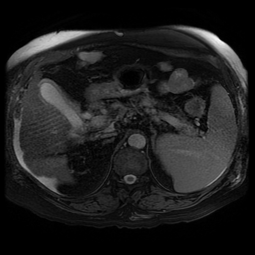
The caput develops due to recanalization of the para-umbilical vein in the falciform ligament, a structure that usually occludes in the second week of life. As with all collateral or variceal vessels identified at CT, MRI or ultrasound it is critical to the diagnosis to consider the direction of flow and what the objective of the varices is. The object of this recanalization is to divert portal venous blood away from the high pressure liver. As the venous vessels radiate from the umbilicus they anastomose with systemic veins diverting portal venous flow. Rarely the inverse direction of flow can be seen in patients with inferior vena cava obstruction. Although recanalization of the portal vein is not uncommon a fully formed Caput medusae is an indication of severe portal hypertension.
More common attempted porto-systemic anastomoses include gastro-oesophageal varices (anastomosing with the systemic lower oesophageal veins) or lienorenal shunts (splenic veins anastomosing with the left renal vein). Portosystemic shunts can also occur around the rectum due to anastomosis of the superior rectal veins (draining to the portal venous system) and inferior rectal/anal veins anastomosing to the internal iliac circulation, although these are also uncommon and seen in very advanced cases.


 RSS Feed
