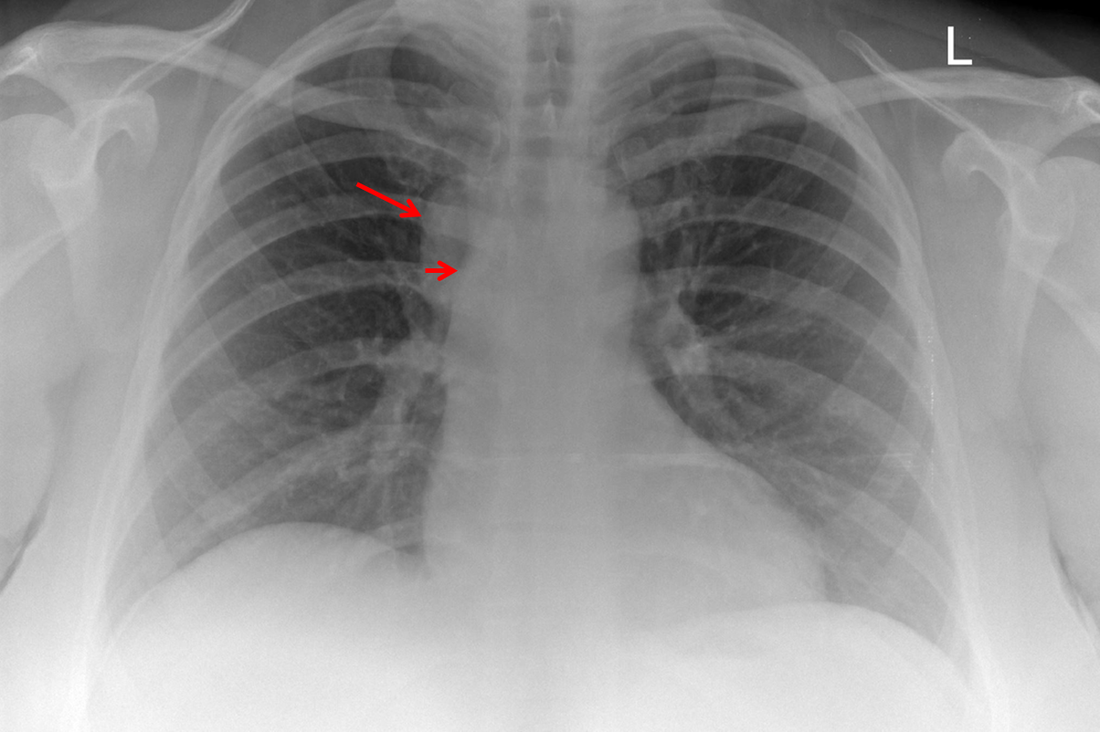
The CT images confirm that there is a mediastinal abnormality that is well defined, homogeneous and of unform relatively low density, less than solid.The abnormality is unilocular and single. The abnormality lies in the middle/posterior mediastinum and as can be confirmed on the coronal reconstructions is separate from the trachea. Hence there is no silhouette sign, with no loss or widening of the right paratracheal stripe.
These appearances are diagnostic of a bronchogenic cyst, also referred to as a foregut duplication cyst. These lesions are very common in this location and more typically right-sided, extending into the azygo-oesophageal region as in this case. Although the density of the abnormality is frequently fluid, the abnormalities may be slightly hyperdense, indicating a slightly proteinaceous contents. Similarly on MRI it is in my experience more typical to see mild T1 hyperintensity rather than the typical low T1 signal characteristics often described. No enhancement is noted within the abnormality on CT or MRI although occasionally mild enhancement of the wall of the cyst can be demonstrated.
Most bronchogenic cysts are asymptomatic discoveries and often measure as much as 10 cm at presentation. Surgery is indicated for comparison symptomatology which is unusual. Secondary infection can be an occasional cause for presentation. The diagnosis should be suspected on plain films where there is a well-defined lesion in a roughly spherical configuration particularly in the middle/posterior mediastinum near the carina.


 RSS Feed
