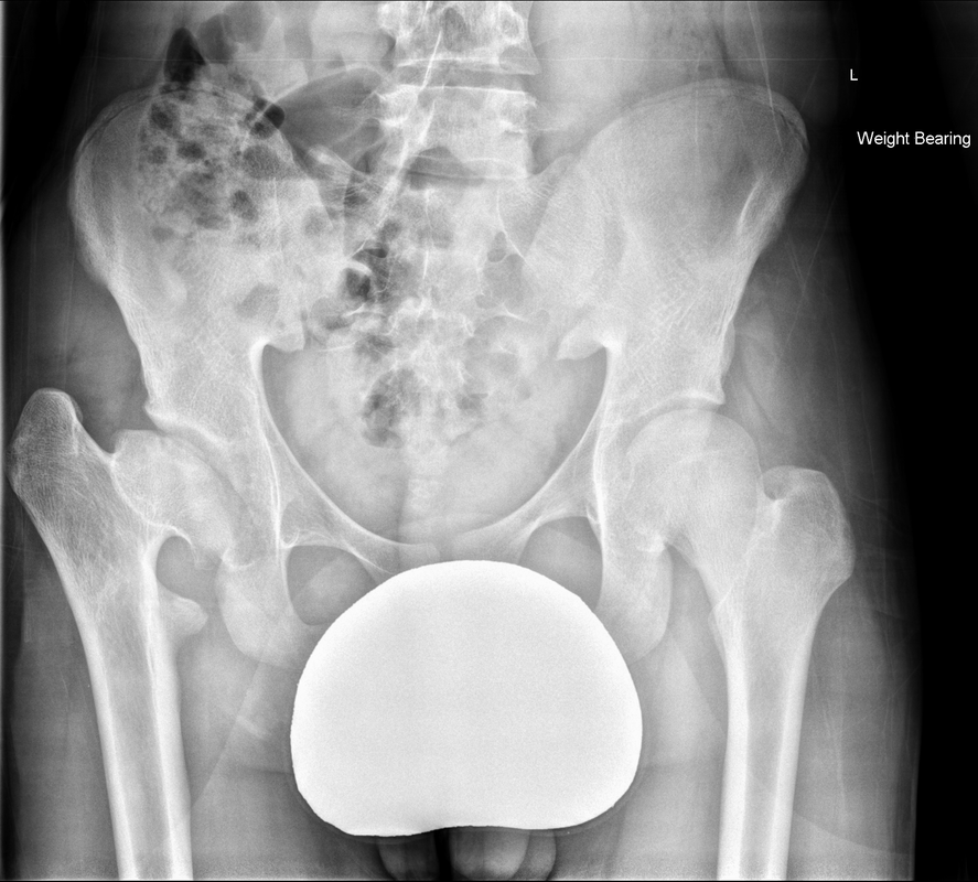
These appearances are highly suggestive of remote missed slipped capital femoral epiphysis (SCFE). SCFE is a diagnosis of prepuberty and puberty typically occurring in the acute phase in children (boys>girls) with a greater incidence in children that are overweight or are suffering from hypothyroidism, hypopituitarism or hyperparathyroidism. Metabolic associated causes may present earlier than 10 years of age and are more likely to be bilateral. Approximately 5-7% of cases have an associated familial predisposition.
The cause of the slipped capital femoral epiphysis is in essence a Salter Harris type I injury. It is thought to occur during periods of rapid growth during which the physis anatomically widens and the biomechanical stresses become more oblique and results in posteromedial slippage.
Most SCFE is picked up acutely, or even in the chronic phase (after 3 weeks of symptoms), however, if missed long term then fusion of the physis can occur in a deformed position. Chondrolysis of the articular surface can occur with premature degeneration of the hip joint. This degeneration typically occurs beyond the fifth decade. This may present as in this case with foreshortening or chronic pain.
A further complication of missed SCFE is appreciated in this case. Look carefully at the right femoral head. It is irregular in shape and mildly sclerotic. There is a small fragment of the femoral head that is separate from the remainder of the femoral head. These are appearances of avascular necrosis. Avascular necrosis is a serious complication of SCFE, and may further predispose to premature degeneration, and a reason why early diagnosis is essential. Avascular necrosis is thought to occur as a result of raised intracapsular pressure in conjunction with kinking of retinacular vessels that is thought to result in hypoxia leading to avascular necrosis. The risk of avascular necrosis appears higher in unstable SCFE (able to walk with our without crutches) compared to stable SCFE (unable to walk).
Treatment both in the acute phase and in the delayed phase id by osteotomy and pinning.


 RSS Feed
