
|
2 day courses:
Mar 20-21 course now fully booked Mar 4-5 course only 3 places left Limited availability for Mar 11-12 and Mar 15-16 1 day written course: Limited availability for Mar 17 Case to Ponder 72: Delayed presentation of missed slipped capital epiphysis with avascular necrosis18/10/2016 Radiology Level: FRCR, ABR, EDiR, Radiology Junior +  This case demonstrates an asymmetrically irregular appearance of the right femoral head and neck. The right femoral neck appears excessively flexed and the right femoral head appears “mushroomed” with a flattened widened appearance. This widened metaphysis is often referred to as coxa magna. The acetabulum is more steep on the right side than on the left, suggesting a developmental cause. In combination the right femoral head is subluxed superiorly. This would result in clinical foreshortening of the right leg.
These appearances are highly suggestive of remote missed slipped capital femoral epiphysis (SCFE). SCFE is a diagnosis of prepuberty and puberty typically occurring in the acute phase in children (boys>girls) with a greater incidence in children that are overweight or are suffering from hypothyroidism, hypopituitarism or hyperparathyroidism. Metabolic associated causes may present earlier than 10 years of age and are more likely to be bilateral. Approximately 5-7% of cases have an associated familial predisposition. The cause of the slipped capital femoral epiphysis is in essence a Salter Harris type I injury. It is thought to occur during periods of rapid growth during which the physis anatomically widens and the biomechanical stresses become more oblique and results in posteromedial slippage. Most SCFE is picked up acutely, or even in the chronic phase (after 3 weeks of symptoms), however, if missed long term then fusion of the physis can occur in a deformed position. Chondrolysis of the articular surface can occur with premature degeneration of the hip joint. This degeneration typically occurs beyond the fifth decade. This may present as in this case with foreshortening or chronic pain. A further complication of missed SCFE is appreciated in this case. Look carefully at the right femoral head. It is irregular in shape and mildly sclerotic. There is a small fragment of the femoral head that is separate from the remainder of the femoral head. These are appearances of avascular necrosis. Avascular necrosis is a serious complication of SCFE, and may further predispose to premature degeneration, and a reason why early diagnosis is essential. Avascular necrosis is thought to occur as a result of raised intracapsular pressure in conjunction with kinking of retinacular vessels that is thought to result in hypoxia leading to avascular necrosis. The risk of avascular necrosis appears higher in unstable SCFE (able to walk with our without crutches) compared to stable SCFE (unable to walk). Treatment both in the acute phase and in the delayed phase id by osteotomy and pinning. "I wanted to thank you for all your help. I have been to other frcr courses but yours is truly very different. You showed a variety of cases (some very difficult ones) and taught me about the pathology and good technique to present cases. None of the other courses do this. Not only did this help me in the exam but in my normal day to day practice.
The written course was useful as you went through the individual sections present in the exam and explained the significance of them in a way no other course does. Overall I feel you were really focused and dedicated in making me pass, the before and after coursework you gave with the feedback was excellent. Thank you for helping me to pass and actually making me a better Radiologist." Exam Scores: Rapids 7, Longs 6.5, Viva 7, Viva 7 More than one previous exam attempt Anonymous Candidate, England Attendee at Original and Written Course Exam Pass "Thank you for all your help and advice, it really helped. I am very grateful. I will recommend your course to everyone taking the exam. You are such a talented and inspirational educator, your trainees are lucky to have you. Your exam technique tips were clearly well above any other 2B course on the market, makes your course unique and essential for passing the exam! I am grateful for the feedback I received from you. It really helped in terms of an exam point of view as I definitely speeded up for the long cases and was more definitive about my diagnosis in the viva." PD, England Previously unsuccesful candidate Attendee at Original Course and Written Course Exam Pass Thank you for your interest and queries.
Only 1 place left for Mar 20-21 2017 2 day course Only 3 places left for Mar 4-5 2017 2 day course Limited availability for Mar 11-12, new date of Mar 15-16 and Mar 17 written course. Radiology Level: FRCR, EDiR, ABR, Radiology Junior + 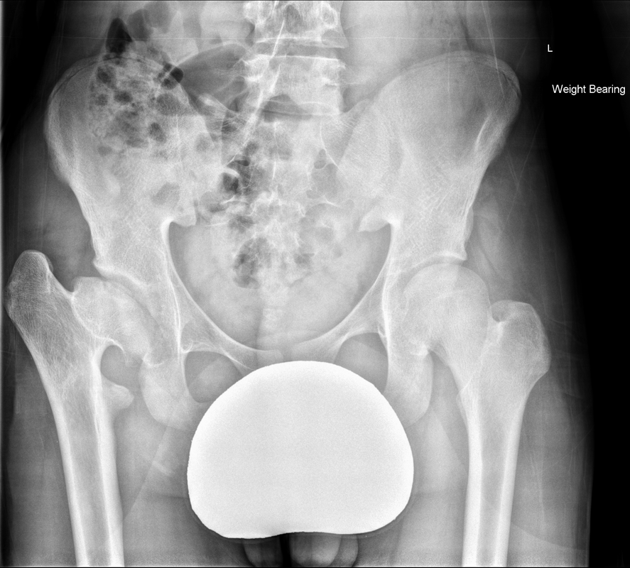 Delighted to be able to offer an additional new date for Original 2 day course on Mar 15-16 2017. Current 2 day slots almost completely full!
The initial chest radiograph demonstrates an abnormal opacity in the right inferior hemithorax. This demonstrates a parallel orientated opacity to the right hemidiaphragm, with some very medial loss of visualisation of the right hemidiaphragm. The abnormality is not anatomically suggestive of a parenchymal abnormality and has vessels seen to be coursing through it, further supporting this is not likely due to a parenchymal air space opacity. There is no blunting of the right costophrenic angle to suggest this is loculated pleural fluid elsewhere. The left hemithorax is clear and the cardiomediastinum a little distorted by a mild scoliosis but otherwise unremarkable. It is noted that there are old posterior fractures of the right ninth and tenth ribs.
The CT examination is illuminating. This demonstrates no overt pleuroparenchymal abnormality. There is elevation of the anterior aspect of the right liver lobe with a notched appearance to the lateral margin of the liver on the bottom left image. Also noted is that there is low attenuation along the vessels in the anterior segment of liver. This suggests that there is segmental mild biliary obstruction in this region, but not centrally in the porta hepatis. The remainder of the right posterior liver and left liver lobe are unremarkable. The right lateral margin of the liver in contact with the right lateral chest wall has lost its normal rounded configuration and has a squared off appearance. These appearances are indicative of a right diaphragmatic traumatic injury with partial herniation of the liver through the diaphragmatic tear. This results in the notch in the lateral liver aspect as well as the segmental ductal obstruction of the herniating segments. Diaphragmatic tearss are not uncommonly delayed presentations following trauma. In part this may because patients with trauma may be treated with positive pressure ventilation which may suppress a diaphragmatic injury. But also tears can enlarge with time becoming sympomatic. Diaphragmatic injuries are commoner on the left side and less so on the right side, thought to be due to the protective of the liver. Most commonly these are due to increased intra-abdominal pressure from blunt injury but can also of course occur with penetrating injuries. Delayed diagnosis results in increased morbidity and mortality, particularly as larger hernias can become more difficult to surgically repair. CT is essential in confirming the diagnosis of diaphragmatic tears, particularly the coronal and sagittal reconstructions, although these are less helpful on the right side due to close approximation of the liver to the diaphragmatic fibres, frequently with no substantial interposing fat. This case was diagnosed based on the abnormal configuration of the right diaphragm with evidence of prior trauma (rib fractures), aided by the focal biliary tract obstruction. Such cases should be differentiated from diaphragmatic eventration which is due to an anteromedial weakness of the diaphragm. In this context the PA chest x-ray would demonstrate loss of the entirety of the right cardiac border near the elevated segment and there would be no evidence of biliary obstruction present. Spring examination:
Written examination: 1st & 2nd April 2016 Oral examination: 3rd April to 7th April 2016 Autumn examination: Written examination: 30th Sept & 1st Oct 2017 Oral examination: 2nd Oct to 6th Oct 2017 RoyalCollege dates and deadlines: https://www.rcr.ac.uk/sites/default/files/cr2b_2017.pdf Delighted to see that courses for Mar 2017 are filling up faster than ever based solely on word of mouth from prior attendees. Sept courses were fully booked so consider booking early. You can now see the comments of prior attendees too:
|
From Grayscale
Latest news about Grayscale Courses, Cases to Ponder and other info Categories
All
Archives
October 2018
|
 RSS Feed